That said if you want to try to drain a dog cyst at home this post offers tips on how to do it carefully and safely. Papillomas are benign but very contagious.

Skin Cysts In Dogs Everything You Need To Know Vet Approved Advice
An epulis is an oral growth that usually forms on the gum tissue around a tooth.
Blood filled cyst dog. Cysts on the surface are often bald and associated with a patch of hair loss. It feels like a round nodule. My dog max a beagle is constantly getting blood fill cysts on his body.
These cysts are round and have a bluish tint. Some dogs develop cysts that are filled with keratin a skin protein. The most common type of cyst contains a gray brown or yellowish granular cheesy material.
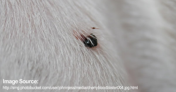
You will either find this extremely gross or extremely awesome if youre the latter- enjoy. It sits on the skins surface or just below the surface. Blood blisters on dogs are usually the result of an injury or trauma of some kind although they can also occur due to a blood clotting problem.
Blood-filled lumps are usually caused by physical trauma. Most are malformations of hair follicles. Follicular cysts appear on or beneath your pets skin.
In the case of an aural hematoma a dog or cat may be itchy as a result of ear mites or an ear infection. Dermoid cysts are rare. Papillomas are warts caused by the papillomavirus.
Interdigital Follicular Cyst on Dogs. The cysts continue coming back just in new locations. Intense scratching and head shaking can inflict trauma to small blood vessels in the outer ear.
False cysts may be formed by hemorrhage or trauma that leads to tissue death. There is no effective treatment. Our vet had the cys.
If a serious infection was involved however the dog will also need to complete a full course of antibiotics which will usually take about two to four weeks. As pressure builds up inside a dogs cyst sometimes a weak spot develops. A sebaceous cyst typically appears as a small raised well-defined round structure in the skin.
A cavity in the skin filled with blood. A Canine skin cyst is an abnormal closed epithelium cellular covering-lined cavity in the body containing liquid or semisolid material. Vascular spaces are separated by fibro-osseous tissue that includes multinucleated cells plump osteoblasts and osteoid formation.
Sebaceous cysts are common in the mouth and chin area of dogs. Sebaceous cysts are common in dogs but unusual in cats with the exception of stud tail on the upper side of the tail. They usually appear as blood- or pus-filled red nodules between the toes usually on the front feet and form as a result of excessive friction or trauma to the webbing between the toes.
A follicular cyst is similar to a blackhead and is more prone to getting infected. Unfortunately draining a cyst on your dog without medical intervention can cause pain while leaving your dogs skin vulnerable to bacterial infection. Dog may have to undergo euthanasia when there is a poor prognosis.
It is either soft or filled with fluid. Your dog can have a cyst anywhere on their body. Follicular cysts are similar to sebaceous cysts and can contain fluid or a thick cottage cheese-like substance.
Diagnosis is with a fine needle aspirate. If the cyst is infected it may. Youll probably find this type of cyst on your dogs head trunk or neck where.
A sebaceous cyst may be firm or it may feel like it is filled with fluid. ABCs have been documented in dogs 6mo-13y that histologically are filled with central blood-filled chambers lined by fibroblasts as opposed to endothelial cells. They can appear on the dogs lips face and inside the mouth.
Such cysts have a hard or solid core. The most common area for you to see a blood blister is. Dermoid cysts are complex congenital cysts that form long before birth.
Blood blisters are hematomas or blisters filled with blood that form under your dogs skin. A Hematoma is usually caused by trauma or injury. If your dog scratches at the bump it may start to bleed.
They can be removed if they cause problems for your dog but in many cases they will resolve on their own. False cysts are fluid-filled structures that do not contain a secretory lining. Hematomas are raised bumps under the skin filled with blood that can form anywhere on your dogs body according to PetMD.
Dog skin cysts can be small and require no medical action removal if they are bothering the dog or removal if diagnosis shows. What Does a Sebaceous Cyst Look Like on a Dog. Fortunately as most cysts tend to occur in the sebaceous glands of the skin the surgical wound will often not be especially deep and will heal in just over a month.
Blood seeps from these vessels and pools into a pocket that forms between the skin and the. Usually these cysts are solitary but some dogs may be prone to getting several cysts in the same area of the body. This is a malignant tumor that can be found internally or as a skin disease.
Carl pops a baseball size sebaceous cyst on a k9 patient. Interdigital follicular cysts in dogs are quite common. Our vet is constantly removing them and draining them.
What Does a Cyst Look Like on a Dog.
If there is a sty on your dogs eyelid it may be very painful. These are common in older dogs and start as small bumps at the margin of the upper and lower eyelids.

Canine Eyelid Masses Acvo Public
Sebaceous Cyst on Dog.

Canine meibomian cyst. Lymphoma Melanoma and Neuroblastoma. Meibomian Cysts also known as eyelid cysts are growths on your dog or cats eyelids. Meibomian gland adenomas are most common.
Cytology and culture of the purulent discharge from one of the meibomian glands yielded Streptococcus species. Cysts may also be present around the mouth. If they become large enough MGAs can cause irritation to the cornea and conjunctiva and may reduce the normal ability to blink.
If they come into contact with the clear surface of the eye the cornea they can cause painful corneal scratches that can lead to corneal ulcers. Simple drainage of the cysts by puncturing them with a 20 gauge cannula is sufficient. A chalazion in dogs is a lump or nodule swelling on the inside edge of the lower or upper eyelid.
Meibomian glands are sebaceous glands that provide an oily secretion to stabilize the tear film over the cornea Common in older dogs meibomian gland tumors are usually benign but a small percentage of them are carcinomas that can metastasize into lymph nodes. Meibomian cysts or tumors occur on or under the eyelid margin. Intervention is only necessary if the cysts touch the cornea and cause irritation.
They can also cause general irritation and inflammation leading to eye infections. Diagnosis of sebaceous adenitis was based on history clinical signs the histological demonstration of multifocal lymphohistiocytic and neutrophilic inflammation targeting the. They secrete an oily substance called sebum which lubricates the skin and hair of animals.
If its a subaceous cyst the bump on your dogs eye may be filled with fluid or solid. Sebaceous cysts appear as growths on the skin of a dog. Bumps in or near the eyes can be uncomfortable because they scratch the cornea or prevent the eye from shutting properly.
Using a wedge resection we removed a large Meibomian cyst from a dogs upper lid. They include nodular hyperplasia sebaceous adenoma sebaceous ductal adenoma sebaceous epithelioma meibomian. A chalazion is also referred to as a meibomian cyst.
Meibomian gland adenoma and adenocarcinomaThese masses arise from Meibomian glands which are specialized glands that line the upper and lower eyelids. A chalazion is also referred to as a meibomian cyst. Older dogs very commonly develop small slowly growing masses on their eyelids.
These pink to pigmented lobulated masses arise from the meibomian glands that line the eyelid margins and may become ulcerated and bleed as they become. In older dogs they tend to develop with no obvious cause and grow slowly over time. Like humans dogs have very tiny oil glands in the deep layers of the skin called sebaceous glands.
The plural form for a chalazion is chalazia. Many of these stay small 2 - 3mm and do not continue to grow further so there is never any rush to have them removed. What causes growths on dogs eyelid.
These are tumours of the meibomian glands of the eyelids. Its unlikely your dog would go blind but if these cysts are left unchecked they could potentially grow large enough to irritate the cornea. Most eyelid tumors in dogs are benign and originate from the glands or skin of the eyelid meibomian gland adenomas epitheliomas melanocytomas and papillomas.
Meibomian cyst is an eyelid lump that is somehow mistaken for a stye. Two-year-old castrated male mixed breed dog with bacterial blepharitis Streptococcus species. Sebaceous adenitis and concurrent meibomian gland dysfunction MGD were diagnosed in a two-year-old mongrel dog presenting with hypotrichosis exfoliative dermatitis and blepharitis.
Note diffuse ulceration of both eyelids with nodule formation crusting and discharge. Cancer of the eye. Meibomian gland adenoma and adenocarcinomaThese masses arise from Meibomian glands which are specialized glands that line the upper and lower eyelids.
Meibomian gland tumors can protrude outward or can extend into the eyelid. Meibomian glands secrete oily substances that help keep the tear film healthy. Meibomian glands secrete oily substances that help keep the tear film healthy.
The top three most common canine eyelid masses are listed below. Removal and Care When Ruptured Bleeding. Resection is usually not possible due to the extent of the cysts.
A combination of oral antimicrobials a tapering dose of steroids and. Initially they are quite small and your dog might not even notice them. Bernard and several Spaniel breeds.
Eyelid tumors can occur in any breed at any age but older dogs tend to present to our service for evaluation. Meibomian Gland Adenomas MGA are benign age related eyelid tumors which result from the accumulation of glandular material. Tumors of the sebaceous and modified sebaceous glands are quite common in dogs.
It is more common in dogs than cats. Canine eyelid masses Eyelid masses or tumors are a common finding in older canine patients although they can occur at any age. The sebum plays a major role in lubricating the hair follicles and hair shafts.
The cysts are filled with honey-colored viscous fluid. Both dog and cat eyes may be affected by these but they are more common in dogs. Masses arising from these glands are often seen protruding from the eyelid.
If you notice a growth on your dogs eyelid it could be whats known as a meibomian gland cyst or chalazion. This is an eyelid condition which is primarily caused by a mere inflammation of the eyelids oil glands meibomian glands. The most common types of tumors appear as neoplasia of the Meibomian gland the primary oil producing glands located in the eyelid margin.
They are usually caused by blocked oil glands. These tumors tend to start out small but usually. Meibomian cyst is also referred as chalazion.
Meibomian gland tumors are tiny slow-growing tumors that form in the meibomian glands of the eyelids. Meibomian gland tumors Meibomian gland adenomas ie benign tumors and adenocarcinomas ie malignant tumors comprise approximately 60 percent of the eyelid tumors seen in older dogs. These masses most commonly arise from the eyelid meibomian glands meibomian gland adenomas or adenocarcinomas or melanocytes melanomas although other cell origins are possible.
Sebaceous glands are microscopic glands found below the skin. The oil produced by these glands is called sebum. It goes without saying that these videos are intended for veterinary prof.
Masses arising from these glands are often.